Quiz Summary
0 of 7 Questions completed
Questions:
Information
You have already completed the quiz before. Hence you can not start it again.
Quiz is loading…
You must sign in or sign up to start the quiz.
You must first complete the following:
Results
Results
0 of 7 Questions answered correctly
Your time:
Time has elapsed
You have reached 0 of 0 point(s), (0)
Earned Point(s): 0 of 0, (0)
0 Essay(s) Pending (Possible Point(s): 0)
Categories
- Not categorized 0%
- 1
- 2
- 3
- 4
- 5
- 6
- 7
- Current
- Review / Skip
- Answered
- Correct
- Incorrect
-
Question 1 of 7
1. Question
1 point(s)
What is the rhythm?
Hint
Look at the rhythm strips at the bottom of the page. Leads II and V5 are shown. The 2nd, 5th, 8th and 11th beats are early. The QRS has the same configuration as the sinus beats so it is supraventricular. There is a P wave prior to every early complex. If you are having trouble finding the P wave in the PAC…it is on top of the T wave preceding the early QRS complex.
-
Question 2 of 7
2. Question
1 point(s)
The QRS duration is ≥ 0.12 sec. Based on the 12-Lead tracing, is there a bundle branch block present?
Hint
The leads to look at are lead I, then V6, and then V1 for confirmation. Look for patterns of bundle branch block in leads I and V6. Then look at V1 for confirmation of the suspected bundle branch block.
-
Question 3 of 7
3. Question
1 point(s)
How do the P waves look?
Hint
Look in lead II and lead V1 for distinctive patterns of atrial hypertrophy. (For a bedside clinician, this not a critical point but it is helpful to start noticing some different configurations of P waves.)
-
Question 4 of 7
4. Question
1 point(s)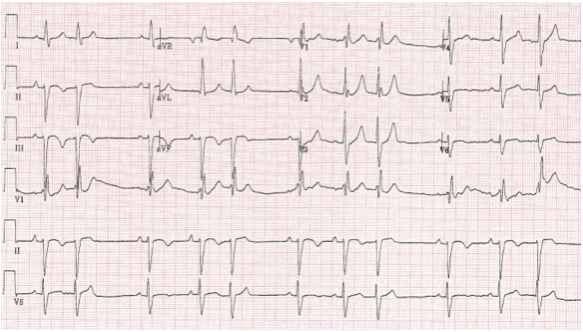
What is the axis?
Hint
Look at lead I and lead aVF. Are they both upright? Do they “point together” with lead I being negative and aVF being positive? Or do they point apart with lead I being positive and lead aVF being negative.
-
Question 5 of 7
5. Question
1 point(s)
Are they having chest pain?
Hint
If the patient is having chest pain, the sequence of examination would be to look for ST segment elevation first. If not present, then look for ST segment depression and/or T wave inversio
-
Question 6 of 7
6. Question
1 point(s)
How do the T waves look?
Hint
Symmetrically inverted T waves indicate ischemia. Asymmetrically inverted are not as suspicious for ischemia.
-
Question 7 of 7
7. Question
1 point(s)
How does the R wave progression look across the precordium?
Hint
V1 should have a small R wave. The R waves will get progressively taller moving toward V6.